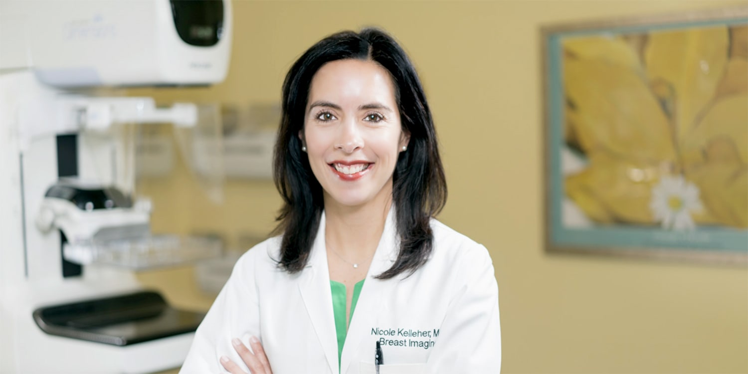
Overview
For most women, breast health is at the forefront of their overall health and wellness. Being proactive about your health includes age and risk-appropriate screening for breast cancer and the early diagnosis of cancer and other breast conditions.
With all the attention around breast cancer, it’s understandable that many women—maybe even you—are fearful of undergoing mammography screening and receiving a diagnosis of cancer. Try to relax. Radiology Associates of Richmond works in hospitals and imaging centers with the most advanced breast imaging technology available for all women—including those at high risk for breast cancer. Our experienced technologists and radiologists are experts in every aspect of breast imaging.
Keep in mind: most breast lumps are NOT cancer and 88 percent of women will NEVER develop breast cancer in their lives—no matter how long they live. Even if you are diagnosed with breast cancer, when you find it early through screening, it is usually treatable.
Symptoms that may prompt a diagnostic mammogram include:
- An abnormal screening mammogram
- A detectable lump in the breast
- Nipple discharge
- Breast pain
- Thickening of the skin on the breast
- Change in the appearance of the breast
Are there any downsides to mammograms?
All medical procedures have some potential risks or harms and no test is perfect. It’s important to understand the pros and cons so you can make an informed decision about whether the benefits of a test or procedure outweigh the risks.
The two major risks of screening with mammography are false positives and false negatives. False positives occur when your mammogram shows an abnormality, but no cancer is present. More than 50 percent of women screened annually for 10 years in the United States will experience a false-positive result. A false negative occurs when your mammogram appears normal; however, you do have breast cancer. Mammograms will miss about 15 percent of breast cancers that are present at the time of screening.
According to the Society of Breast Imaging (SBI), the harms of screening are negligible compared to dying from breast cancer. For every 1,000 women screened, 100 are called back. Of the 100, 81 are negative and rescreened in a year, or have another imaging study in six months. Nineteen undergo a minimally invasive needle biopsy and five of them are diagnosed with breast cancer.
The benefit of mammography, of course, is that early detection of breast cancer means it is less likely to have spread and is easier to treat. We know that the risk of breast cancer increases with age and the benefits of finding cancers early decrease the treatment for breast cancer.
Radiology Associates of Richmond at Henrico Doctors Hospital—Forest and Johnston-Willis are Breast Imaging Centers of Excellence (a designation from the American College of Radiology ) for mammography, breast ultrasound, breast MRI, and stereotactic guided biopsy,. We are also accredited by the National Accreditation Program for Breast Centers.
What happens during a mammogram?
A radiology technologist will place each breast on a special platform on the mammography unit and compress it while she takes two x-ray images. Each x-ray provides a different view of the breast. At that point, the technologist will check the images to make sure they are good quality. Occasionally, we may take a repeat view if you accidentally moved or there are artifacts (for example, a stray hair in the image) that make it difficult to see everything clearly.
The compression might be a bit uncomfortable, but should not be painful. Tell your technologist if it is and she’ll try to make you more comfortable. At Radiology Associates of Richmond, we use the lowest dose of radiation possible while still producing the highest quality image.
TIP Schedule your mammogram for the end of your menstrual period when your breasts are less tender. Cutting back on caffeine for a few days before your mammogram may also help lessen any discomfort from the compression.
What happens after your mammogram?
After your screening mammogram, one of our experienced radiologists will read your x-ray. If your mammogram is normal, we will send you a letter, usually within one to two days.
If something looks suspicious, we will call you within 1-2 days. Only about 10 percent of women who have a screening mammogram are called for a follow-up appointment. We may request a spot compression or additional views, or an ultrasound to evaluate a mass and determine if it’s solid or filled with fluid. If we have prior mammograms, we will compare the current radiograph to earlier screenings. If you’ve had a mammogram at another imaging center, please ask that center to send us your images.
We know it’s stressful to be called back for additional tests. Rest assured, if we ask you to return, a breast radiologist will review your results with you in detail the same day. You won’t have to wait for a call or a letter.
When a traditional mammogram is not enough
For women who are at high risk for breast cancer—for example, those who have dense breasts—or who can’t undergo a mammogram (for example, because they are pregnant), Radiology Associates of Richmond works in hospitals and imaging centers that offer specialty breast imaging technology. These technologies don’t replace traditional mammography, but rather provide additional information when needed.
40% of American women have dense breasts, which makes it difficult to see changes not detectable by traditional mammography
Who Should be Screened?
Screening recommendations vary somewhat based on age and individual risk factors for breast cancer (for example, family history). The Society of Breast Imaging (SBI) and the American College of Radiology (ACR) recommend annual mammograms for women at average risk of breast cancer beginning at age 40 and continuing as long as a woman is living a relatively normal life with a life expectancy of five or more years.
Women at higher risk for breast cancer should talk to their doctor about when to begin screening, which tests are appropriate and how often to screen. The SBI and the ACR considers the following women to be at increased risk:
- Women with certain BRCA1 or BRCA2 mutations or who are untested but have first-degree relatives (mothers, sisters, or daughters) who know they have BRCA mutations
- Women with a 20 percent or greater lifetime risk for breast cancer, based on family history (both maternal and paternal)
- Women with mothers or sisters with pre-menopausal breast cancer
- Women with histories of mantle radiation (usually for Hodgkin’s disease) received between the ages of 10 and 30
- Women with biopsy-proven lobular neoplasia (lobular carcinoma in situ and atypical lobular hyperplasia), atypical ductal hyperplasia (ADH), ductal carcinoma in situ (DCIS), invasive breast cancer or ovarian cancer
